

Pancreatic Pseudocysts: When Is Surgery Necessary and When Is It Not
The human body possesses a remarkable ability to wall off trouble, but sometimes those defensive measures create a whole new medical challenge. When the pancreas experiences a severe bout of inflammation, it can leave behind fluid collections that do not simply vanish on their own. These fluid structures are known as pancreatic pseudocysts. Despite what the name implies, a false cyst is not an insignificant issue. It is a complex condition that requires careful clinical evaluation.
A frequent source of anxiety for many patients is the assumption that diagnosing a pseudocyst automatically means going under the knife. However, modern medical practices emphasize a highly tailored approach. While some pseudocysts resolve quietly over time, others escalate to a point where intervention becomes absolutely vital.
What Exactly is a Pancreatic Pseudocyst
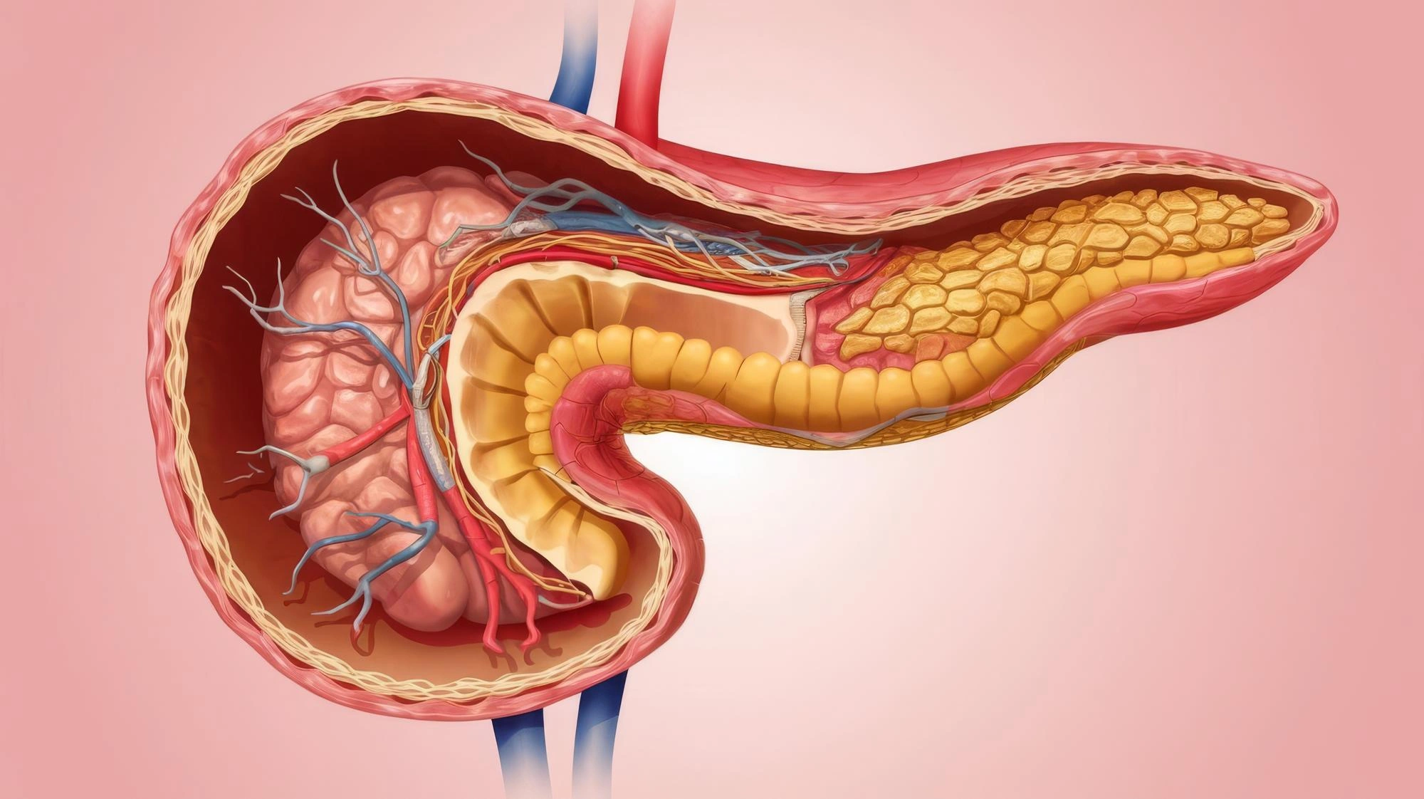
To understand how a pseudocyst behaves, it is helpful to contrast it with a true biological cyst. A true cyst is lined by a distinct layer of epithelial cells that actively secrete fluid. A pancreatic pseudocyst does not have this cellular lining. Instead, it is a localized collection of fluid, pancreatic enzymes, and dead tissue debris enclosed by a thick wall of fibrous or inflammatory tissue.
These structures typically develop as a direct consequence of acute or chronic pancreatitis. When the pancreas becomes inflamed, its delicate ducts can leak enzyme-rich fluids. In response, the body attempts to contain the irritating fluid by forming a fibrous barrier around it. This protective walling-off process usually takes about four to six weeks to fully mature.
The Watchful Waiting Phase: When Surgery Is Not Needed
Not every pseudocyst requires active medical intervention. In fact, a significant portion of these fluid collections will resolve completely on their own through natural resorption. If a patient is completely asymptomatic, a primary policy of careful observation is generally the safest course of action.
During this conservative management phase, several factors influence the decision to hold off on treatment. Cysts that measure less than six centimeters in diameter have a substantially higher likelihood of disappearing on their own. Additionally, if the patient is not experiencing persistent abdominal pain, early fullness after eating, or nausea, there is no immediate pressure to intervene.
As long as regular imaging shows no signs of infection, rapid expansion, or compression of surrounding organs, watchful waiting remains the standard choice. Medical teams track these cysts using regular CT scans or MRI scans to monitor changes in size and wall thickness over several months.
Crossing the Line: When Intervention Becomes Necessary
While observation is ideal, certain clinical triggers indicate that a pseudocyst has become a dangerous liability. Leaving a problematic cyst untreated can lead to severe, life-threatening scenarios.
Intervention becomes necessary when specific red flags appear. As a pseudocyst grows, it can cause significant pressure within the abdomen. Patients often experience a dull, constant ache in the upper abdomen that can radiate to the back, severely impacting their daily quality of life.
Furthermore, the stomach and intestine sit in close proximity to the pancreas. A large, expanding pseudocyst can push directly against the stomach wall, leading to a blockage. This manifests as an inability to tolerate solid foods, persistent vomiting, and rapid weight loss.
How Specialized Scans Dictate the Path Forward
Before any medical procedure is planned, the internal architecture of the abdomen must be mapped out precisely. Selecting the right treatment relies entirely on detailed diagnostic imaging.
Medical experts look closely at how close the pseudocyst sits to the stomach wall. The thickness and maturity of the cyst wall are equally critical because a fragile, immature wall cannot safely hold internal sutures or stents. Advanced imaging also tracks the exact distance between the stomach and the cyst, while distinguishing whether the interior contains pure, free-flowing fluid or is cluttered with solid, damaged tissue debris.
Modern Drainage Techniques: Three Paths to Resolution
If a pseudocyst meets the criteria for drainage, the medical team has several modern therapeutic avenues available. The days of automatically performing open abdominal surgery are long gone, replaced by precise, minimally invasive options.
Conventional Open Surgery
While less common today, conventional open surgery remains a vital backup option. In cases where the cyst wall is unusually thick, or if there is extensive, solid tissue debris that cannot pass through a minor opening, an open surgery called a cystogastrostomy is performed. The surgeon creates a direct surgical connection between the back wall of the stomach and the front wall of the cyst, allowing the contents to drain cleanly into the stomach to be digested naturally.
Laparoscopic Drainage
Laparoscopy brings a minimally invasive approach to this anatomical challenge. Using tiny, keyhole incisions in the abdomen, the surgeon inserts a camera and specialized instruments. They navigate inside, access the stomach, and create the same protective drainage channel into the gastric cavity. This technique significantly reduces postoperative pain, shortens hospital stays, and accelerates the patient's overall recovery timeline.
Advanced Robotic Intervention
Representing the cutting edge of surgical care, robotic-assisted surgery provides unmatched precision. The robotic system allows surgeons to operate with enhanced three-dimensional visualization and highly articulated instruments that mimic the human wrist. This level of dexterity is particularly beneficial when creating precise connections between the stomach and the cyst wall in tight anatomical spaces, minimizing trauma to the surrounding healthy pancreatic tissue.
Comprehensive Care at Jyoti Hospital
Navigating a complex gastrointestinal condition requires an experienced medical team. Located on Thiruvottiyur High Road in Old Washermenpet, Chennai, Jyoti Hospital has stood as a trusted multispecialty healthcare institution for over twenty-five years. Our state-of-the-art facility is equipped with advanced diagnostic imaging suites, modern operation theaters, and dedicated intensive care units designed around absolute patient safety.
The core foundation of Jyoti Hospital is our highly experienced team of specialists and surgeons, many of whom possess more than twenty-five years of individual clinical expertise. By combining sophisticated, minimally invasive techniques, including laparoscopy and robotic-assisted interventions, with a deeply compassionate approach, we ensure that advanced gastrointestinal care remains accessible, safe, and highly effective for our community. Your recovery, comfort, and long-term well-being are the absolute center of our practice.